![<?echo $_SERVER['SERVER_NAME'];?>](/template/twentyseventeen/skin/images/header.jpg)
Abstract Plum blossoms—a wonderful representation of one-dimensional nanorods and three-dimensional nanoparticles under a scanning electron microscope at a magnification of 15,000 times. Liu Ziwei, a Shanghai Institute of Ceramics, Chinese Academy of Sciences, photographed the fruit - fluorescent staining of rat brain slices. ...
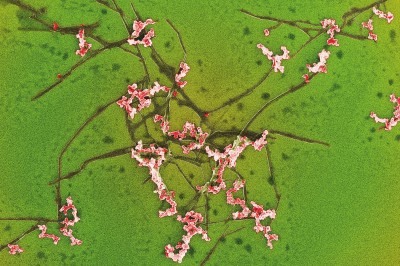
Plum blossoms - a wonderful representation of one-dimensional nanorods and three-dimensional nanoparticles at a magnification of 15,000 times scanning electron microscopy. Photograph by Liu Ziwei, Shanghai Institute of Ceramics, Chinese Academy of Sciences
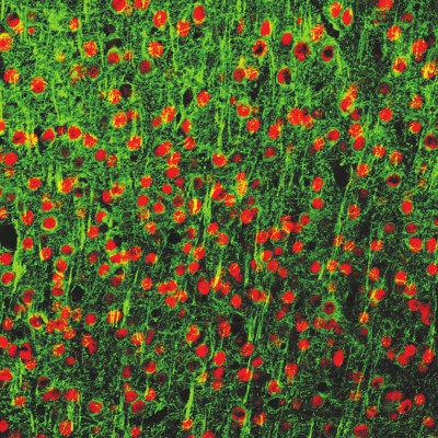
Fruits are plentiful - fluorescent staining of rat brain slices. Zhang Lei, Shanghai Institute of Materia Medica, Chinese Academy of Sciences
The poison of the heart - the small molecule of the drug causes cardiotoxicity to the potassium channel block. Photograph by Liu Lili, Shanghai Institute of Materia Medica, Chinese Academy of Sciences
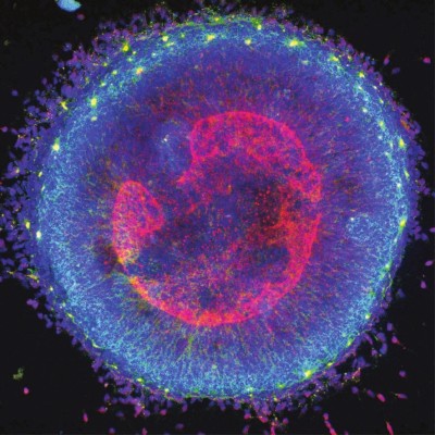
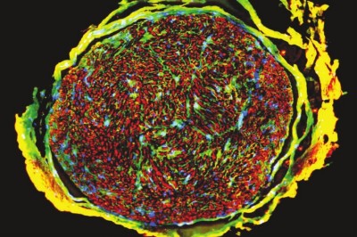
Dark night alum - immunofluorescence staining of human embryonic stem cells during neuronal differentiation to form a Rosset (rose cluster structure). Chen Hao, Shanghai Academy of Sciences, Chinese Academy of Sciences
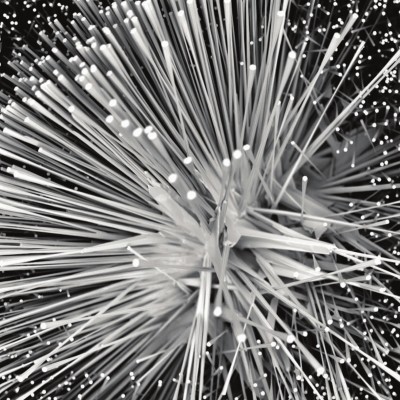
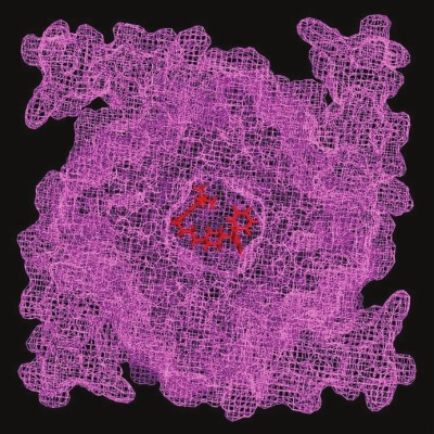
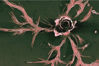
A sharp rim – a zinc oxide whisker observed at 3,550 times. Photograph by Lin Zebin, Fujian Institute of Physical Structure, Chinese Academy of Sciences
Blooming—The flower-like oxide formed on the surface of the TiAlN/SiN composite film after high temperature oxidation is dyed and shaped like a blooming rose. Wang Ruishe, Ningbo Institute of Materials, Chinese Academy of Sciences
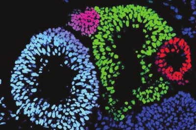
Garland - a neuroepithelial cell induced by human embryonic stem cells that exhibits a floral ring structure under an optical platform confocal microscope. Shanghai Institute of Bioscience, Chinese Academy of Sciences
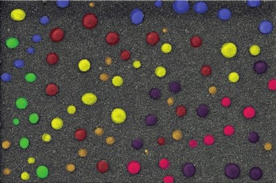
Colorful pebbles - copper-chromium metal co-doped diamond-like carbon films, prepared by PVD (physical vapor deposition) technology, sprayed with platinum. Photograph by Sun Lili, Ningbo Institute of Materials, Chinese Academy of Sciences
Psychedelic - a three-color fluorescent marker of the transverse section of the human ulnar nerve root. Sun Peng, Shanghai Institute of Materia Medica, Chinese Academy of Sciences
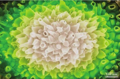
Flower language - the state of gold nanoparticles exhibited during SEM testing. Photograph by Gu Jincui, Ningbo Institute of Materials, Chinese Academy of Sciences
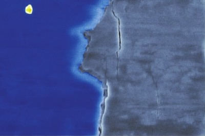
Looking at the moon, the TiAlN/SiN composite film is cracked after the scratcher has been scratched over the surface, and it looks like a person's side face after dyeing. The moonlight night triggers homesickness and is meaningful. Wang Ruishe, Ningbo Institute of Materials, Chinese Academy of Sciences
Who can believe that the plum blossoms in front of this "shadowing and fragrant" are actually scientists taking one-dimensional nanorods with a diameter of about 100nm and nanoparticles of 200-400nm, which are "red" under the scanning electron microscope with a magnification of 15000 times. A special image of "green". Scientists not only have the ability to explore the unknown, but also have a pair of unique eyes. This group of photomicrographs in this edition are from the hands of scientists in various disciplines of the Chinese Academy of Sciences. They capture the “by-products†of these scientific studies through microscope observation. Although these wonderful images also contain a number of "photography" elements, they still differ from the traditional principles of photography.
Microphotography is a technique that adds imaging to the microscopic technology, which is a technique that uses professional microscopic equipment to display the field of view under the microscope as a photo. With the rapid development of modern computer technology, today's photomicrography eliminates the need for camera-controlled shooting. Instead, it combines advanced optical microscope technology, photoelectric conversion technology, and liquid crystal screen technology into a scanning electron microscope instrument to record the observed objects. The fine structure of the image is much higher and sharper than before. This has become an indispensable technology for the research work of scientific research, teaching and medical science and technology personnel.
Appreciating the micro-photographs captured by scientists with Goggles, people can't help but admire: Science can be so beautiful!
The work "Bloom" is a flower-like oxide formed on the surface of TiAIN/SiN (a new type of coating material) composite film after high temperature oxidation. The shape looks like a beautiful rose; "Flower" reflects that gold nanoparticles have many special The physical properties, the author's stunning picture in the electron microscopy test, does not lose to the nature of the red and green; "Feng Man Bi Lu" through the 3550 times observed in the form of zinc oxide whisker, the painting presents each whisker Made up of four needle-like crystal fibers, like a hedgehog with a needle coat; "Psychedelic" comes from the three-color fluorescent marker of the transverse section of the human ulnar nerve root, red is synaptic protein, green is myelin, blue The color is the nucleus, much like the fireball of the cosmic explosion; "The poison of the heart" shows the peculiar sight of the small molecule molecule on the cardiotoxicity caused by potassium channel blockade...
Microphotography, as a means of scientific research, truly represents the fine structure and morphology of various objects, and is the basis for the recording and analysis of scientific research results; they not only reflect the value of scientific micro-world research, but also bring extraordinary beauty.
Visiting the spring flowers, stopping in the garden flower gardens, is also in the fascinating science laboratory.
Cold Drawn Pipe,Seamless Duplex Tube,Cold Drawn Steel Tube,Hot Rolled Seamless Steel Pipe
Zhejiang Ruimai Stainless steel Tube Co.,Ltd. , https://www.ruimaitube.com